Moles
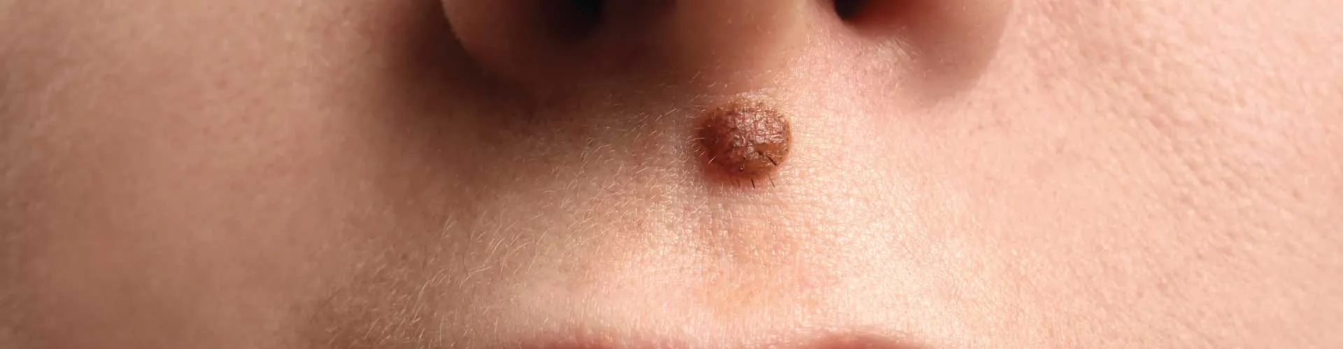
A mole, the medical term is ‘nevi’, the most common pigmented skin growth, appear as small raised dark brown spot, though the skin color can range from pink to black. The pigment-producing skin cells melanocytes are evenly spread throughout our skin, though they can grow in clusters thus forming moles.
Moles can exist at birth or appear later. Generally, they appear during childhood and adolescence. Most adults have 10 to 40 moles.
Genetic predisposition and abnormal exposure to the sun play an important role. Hormonal changes during puberty and pregnant women can provoke melanocyte to develop moles.
Most moles are harmless, but in rare cases they become cancerous. However, monitoring of the existing moles and sudden multiple new eruptions is an important step detecting skin cancer, called malignant melanoma.
Most moles do not require treatment except for those cosmetically undesired, but some cases require removal of the mole.
Types of Moles
Congenital
Congenital moles are present at birth and are caused by melanocyte cells in the dermis-epidermis. These moles vary in size from small, medium, or giant. The giant congenital mole can turn cancerous as they ages to adult life.
Acquired mole or Common mole
Acquired moles are those that you develop later in life. Most of these are brown to dark brown and are found in people regularly exposed to the sun. These are dome-shaped smooth, pigmented with a distinct shape, and 3-6 mm in diameter. These moles are less likely or not likely to turn into cancer. Mostly they develop lighter skin since they have lower levels of melanin.
Acquired moles or nevi are further differentiated depending on the location of involvement:
- Junctional Melanocytic Nevi: : Here the melanocytes accumulate or proliferate between the epidermis and dermis to form these moles. They are raised, uniformly pigmented with a diameter of 2-6mm, and have regular borders.
- Compound Nevi: Here the melanocyte proliferates in the epidermis and migrates into the dermis with the maturation of the cells in the deep dermis. They are round or oval with slightly raised in the central part and have flattened distinct borders.
- Intradermal Nevi: They are located in the dermis and proliferate in deeper skin; hence they are not as pigmented as junctional or compound nevi. They are formed as we age and are flesh-colored, dome-shaped papule or nodule.
- Atypical mole: also called a dysplastic nevus. Atypical moles are often odd or irregular shapes, larger than a pencil end or eraser, and appear blurry with mixed color (brown, red, and pink). They are either raised or flat and look like melanoma. However, it is not melanoma but has a higher risk of forming melanoma or cancer. They can appear anywhere on the body, often seen on the trunk. Higher risk of getting melanoma with atypical mole:
- Four or more atypical moles.
- History of having melanoma
- First-degree relative or parents having melanoma ( brother, sister, or child)
Let us know how to differentiate a ‘regular mole’ from an irregular mole or dysplastic or cancerous mole
| Regular mole or Benign mole |
Dysplastic or Cancerous mole |
| Appear anywhere on your skin & below the age of 20 |
Appear anywhere on your skin, preferably trunk & after 30 |
| Smooth surface and asymptomatic |
Associated with itch or ooze or pain |
| Dark brown or black |
Mixed color - brown, red, and pink |
| Symmetrical in shape |
Asymmetric shape |
| Regular Border and or distinct |
Irregular border or ill-defined border |
| Evenly pigmented |
Uneven pigmentation |
| Size 2-6mm |
Size exceeding 6mm |
| Darken during puberty, pregnancy or with time |
Evolving or change in shape, size, surface, and color |
| Usually harmless & does not require removal, except cosmetic desire to remove |
Careful monitoring and need early intervention to remove |
The above table differentiates the malignant change in a mole and a need for the doctor visit. Most moles are benign and harmless. Older people have a higher risk. Unusual changes in a mole or pigmented spots as ages into adulthood need for a check-up or doctor visit.
The main risk factor with a mole is Melanoma, a type of skin cancer developing from a mole.
Let us know a few high risks of getting melanoma in a mole are:
- Congenital mole: Born with large size mole > 5 cms, could potentially turn cancerous as the child ages in early adulthood.
- Atypical or dysplastic nevi or irregular mole: Having unusual-looking moles with a dark brown centre and lighter uneven borders and even called ‘halo moles’ due to its characteristic appearance. Few types of atypical moles are genetically predisposed to form melanoma
- Large number of moles: Having more than 50 regular moles on your body or 20 moles on their arms, are prone to develop malignant melanoma and must take preventive measures. The higher numbers of moles in a woman have a direct link with the risk of breast cancer.
However, people born with several moles or unusual looking moles or large mole at birth have a higher risk of melanoma.
Let us know a few of the preventive measures in higher-risk groups:
- Sun protection
- Physical by using wide-brimmed hats, and protective clothing (long sleeves or pants)
- Chemical by using water-proof sunscreens with SPF 50+
- Avoid overexposure to the sun between 9 a.m. to 4 p.m.
- Regular examination of moles and sun-exposed skin on a monthly or quarter yearly basis, as of risk factors.
- Avoid suntan parlor
- Intervention treatment either skin biopsy or removal of a mole is required for the following conditions:Most moles do not require skin biopsy and or removal.
- Cosmetic undesired normal moles can be removed with the biopsy punch or surgical excision or radiofrequency device or CO2 laser.
- Suspicious (could be skin cancer) like abnormal or atypical moles are not malignant, and may or may not need to be removed. If removed, proper stitches to be done and regular check-ups are required.
- Malignant moles or melanoma: These are cancerous moles and must be removed immediately. Surgical excision has to be performed.