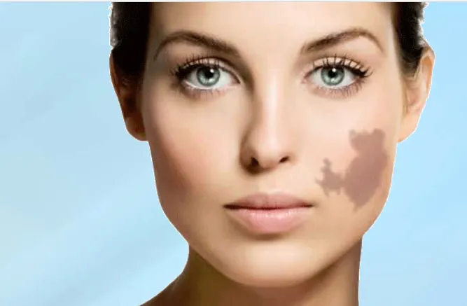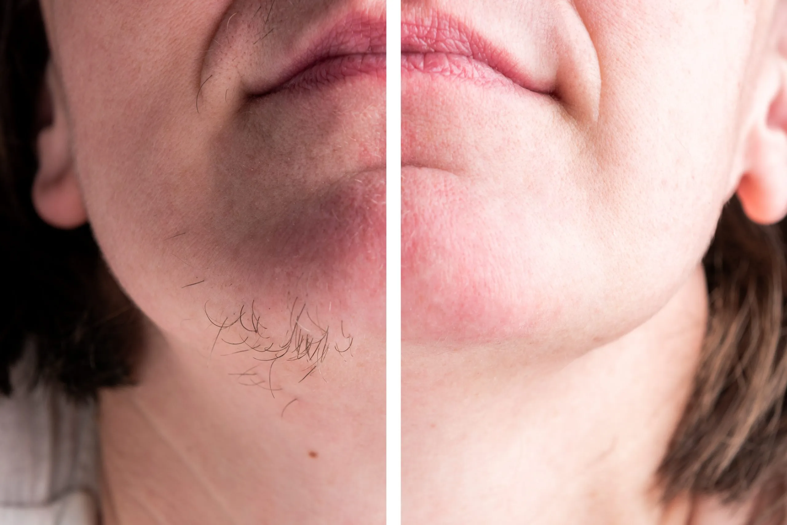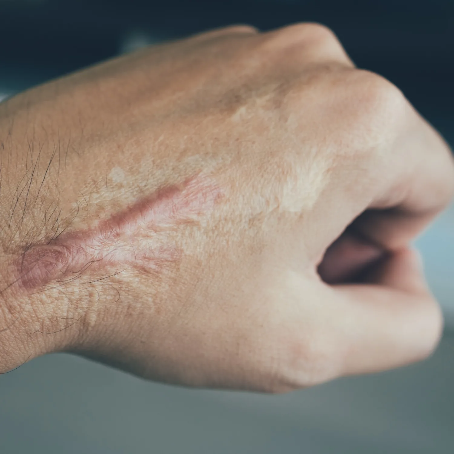
Keloid scars are thick clusters of scar tissue that and extend beyond the edges of an original wound or trauma. Most keloids are raised, round, or oval with regular margins and some with irregular margins have a crab claw-like look. Early lesions are red with soft or doughy consistency and mature or older ones have are brownish red with rubbery or hard and become wider over time. Keloids can occur anywhere on the body but occur more commonly with little underlying fatty tissue such as the face (low part), neck, ears, chest, shoulders, upper arm. They are often associated with itching, pain, anxiety or depression, and disruption of daily activities.
Hypertrophic scars are similar, slightly smaller than keloid, develop directly at a wound site, and do not go beyond the bounds of injury. They are often raised, red, and regress over time.
Keloids & hypertrophic scars are raised to have a heaped-up appearance. These scars rise above the rest of the skin; keloid remain elevated above 4mm to the skin surface
| DO | DON'T |
|---|---|
| Do consult the expert skin professional treating the same. | Do not expose to sun |
| Do use a broad spectrum sunscreen daily | Do not go for ear or body piercing and tattoos |
| Do use a protective clothing on outdoor to protect the scar | Don’t pop pimples |
| Do be careful while shaving to prevent nicks and cuts | Do not scrub or scratch your skin too roughly |
| Do treat the injuries and burns immediately | Do not ignore at initial stage of formation |
| Do take care if you have acne and maintain a proper skincare regimen | Do not go for cosmetic surgery, if family or in personal have keliod tendency |
| Do massage the scar | Do not smoke, as it decreases blood circulation in the skin and inadequate wound healing and possibly tissue loss |
| Do keep the wound moisturized | Do not have un-realistic expectations from the treatment |
The exact formation of keloid is still not known. They are formed due to an overactive healing response. After any injury to the skin, it forms a gap. To bridge the gap our body produces granulation tissue over the injured area, predominantly consisting of fibrous protein or collagen. Usually, the body knows when to stop this process of healing, but in some people genetically pre-disposed there is an imbalance between collagen stimulators and inhibitors lead to the formation of keloid. The thick clusters of collagen are formed leading to scar tissue that continues to grow in all directions, involving the surrounding skin of the original site of a wound.
Hypertrophic scars are similar, but are confined to the wound borders and usually regress over time. This scar usually appears within a month of injury, in comparison to keloids may take three months or even years to develop. Both represent abnormal fibrotic responses to dermal injury, with exuberant deposition of collagen, but an equilibrium stage reached once a thick layer is formed after approximately 1.5 to 2 months; and overtime elastic fibers are formed.
| Hypertrophic scar | Keloid |
|---|---|
| Remain confined to border of the original wound | Extend beyond the border of the original wound |
| Mostly asymptomatic | Associated with itching or if overlying a joint can restrict movement |
| Soft to a firm inconsistency | Firm to a hard consistency |
| Arise in any location; commonly occur on extensor surfaces of joints | Commonly occur on the sternal skin, shoulders and upper arms, earlobes, and cheeks |
| Appear within one month | Appear at three months or later grow for years |
| Flatten spontaneously in time | Remain elevated more than 4 mm |
| Regress with time | Grow for years |
| Less association with skin color | More common in darker skin types |
| Respond well to treatment | Resistant to treatment |
| May consist of a hair follicle and adnexal glands | Devoid of hair follicles and adnexal glands |
The treatments for Keloid and hypertrophic scars are similar, but hypertrophic scars have a better prognosis with age. Hypertrophic scars can easily be treated however keloids are resistant to treatment. Treatment is based upon the depth, size, and location of keloid. History of previous treatment and its response is important before starting treatment. There are different treatments available are mentioned below:
Any physically healthy, mature, and realistic expectations with a desire to reduce or remove a keloid scar is an ideal person.
Topical Silicone gel or creams or sheets are useful to some extent.
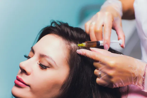
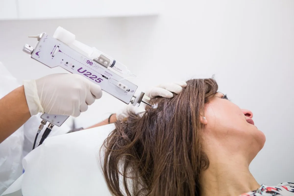
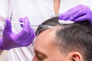
Intralesional steroid or 5FU or verapamil injections are often used to stop the growth of keloids. Injecting long-acting cortisone (steroid) into the keloid once a month is the most effective first-hand treatment.
Retinoic acid, Interferon (IFN) therapy, doxorubicin, bleomycin, imiquimod 5% cream, tamoxifen, and tacrolimus
Antiangiogenic factors, including vascular endothelial growth factor (VEGF) inhibitors (eg, bevacizumab),
Photodynamic therapy, UVA-1 therapy, transforming growth factor (TGF)–beta inhibitors, tumor necrosis factor (TNF)-etanercept, which are all directed at decreasing collagen synthesis.
Compression therapy or Pressure garments it suppresses collagen production
Occlusive scar dressings should be worn for at least two to three months and can be extended as per requirement.
Treat secondary infection in time, drain the abscess and sinus, and remove coiled hair inside the keloid.
Cryosurgery: most commonly used for small or limited keloids, most suitable and is often used parallel to steroid injections.
Laser therapy; very good at improving skin texture and color, and in combination with other treatments flatten out the keloid.
Pulsed Dye Laser: most effective if used early and in combination with other techniques. The principal effect of a pulsed dye laser is on scar microvasculature, reducing erythema and pruritus, and improving skin texture and significant improvement in the scar. Fractional Co2 creates deep channels into thick scar tissue with debulking mode; and if used in combination collagen remodeling agents can almost flatten the hypertrophic scar and keloids, if used for few consecutive months.
Laser hair removal: hair follicle and sebum production act as a trigger; hence laser hair removal can be added Fractional MNRF and individual radiofrequency devices in combination with cryo and intralesional steroids give excellent results.
Enerjet: Hyaluronic acid (HA) is a glycosaminoglycan that plays an important role in the reorganization of the extracellular matrix (ECM) during the skin wound healing process. Decreased presence of HA is a major characteristic of keloid and hypertrophic scarring
Enerjet uses Jet Volumetric Remodeling (JVR) technology a high-pressure jet technology to laterally introduce healing agents deep into the dermal layer of the skin for the reduction of keloid.
Surgical excision: Surgical Removal is can be done for larger, more extensive, and especially over the ears. The technique involved may employ a scalpel, radiofrequency device, or CO2 laser removal. It’s better to avoid surgical excision as it may result in a new keloid, and even larger than the original one.
Radiation therapy: carried out soon after surgery
We at Clear Skin laser center provide all the above mentioned, advance, and evidence-based laser treatment for keloid treatment. Treatment protocol for the management of these scars will extend from 6 months to 2 years; once in a month as out-patient based sessions.